The longer one suffers from diabetes the greater risk there is of developing retinopathy.
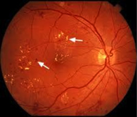
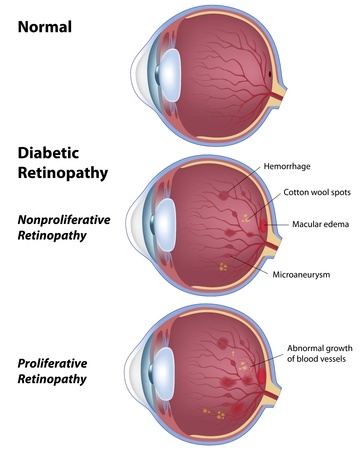
The first pathological change occurs in the smallest retinal blood vessels called “capillaries”. This is where oxygen exchange occurs. The capillary wall develops a weakness and results in a bulge called a “microaneurysm”. Plasma leaks and bleeding can occur. This stage is called background retinopathy. If this occurs at the center of the retina, it is called maculopathy and vision is reduced

Dr. Matzkin can diagnose diabetic retinopathy with a clinical exam, photographs and a fluorescein angiogram where a dye injected into the vein can highlight leaking and abnormal retinal blood vessels.


